I think my model is not having good precision because I have not have segmented the tumor in MRI images. You see MRI image file in .nii have many slices in it and I am not sure whether the image reader is reading it in 3D or not as I can only see one slice of image.
I think I will need to segment the tumor either manually placing ROI or semi-automatically. Is there any way I can achieve this using Knime?
I want to know is it possible to run ANTs in Knime or similar tool like that. http://stnava.github.io/ANTs/
If not can one create a node? How it takes to create a node.
Is there any alternative for achieving this image segmentation, maybe ImageJ? (Find edge node in ImageJ?)
Also is it possible to know whether the images I am importing in Image reader node are 2D or 3D stack like in MRI image which will have slices e.g. 28 etc.
@christian.dietz
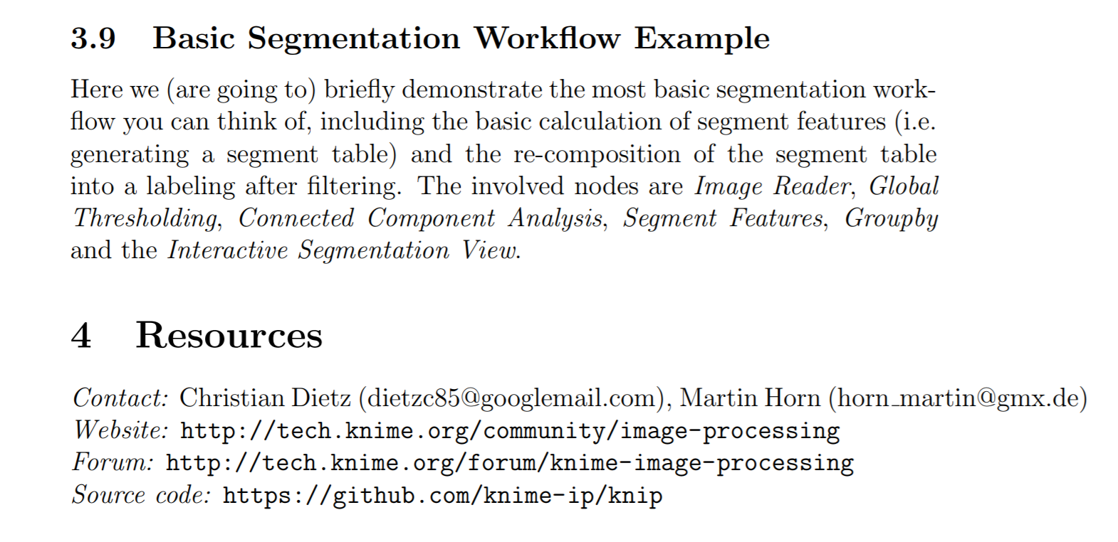
I have found this image post processing manual for image segmentation.
But how to apply this segmentation and thresholding in 3D stack image.
Also I still could not figure out how to scroll them in KNIME image reader node.
maybe it’s worth reading “KNIME for Open-Source BioImage Analysis - A Tutorial” (you can find it via scholar.google.com). This tutorial explains the basic concepts of KNIME Image Processing. Also, please find several tutorials and example workflows here Image Processing | KNIME.